Home

Si verificano Biscotto fenomeno verocay bodies vincere footpad labbra

Verocay bodies | Radiology Reference Article | Radiopaedia.org

CNS Pathology
CNS Pathology

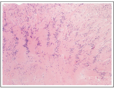
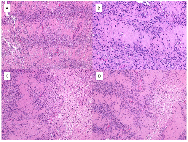
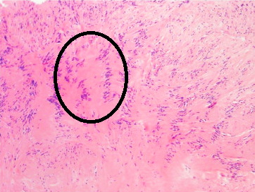
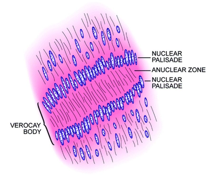
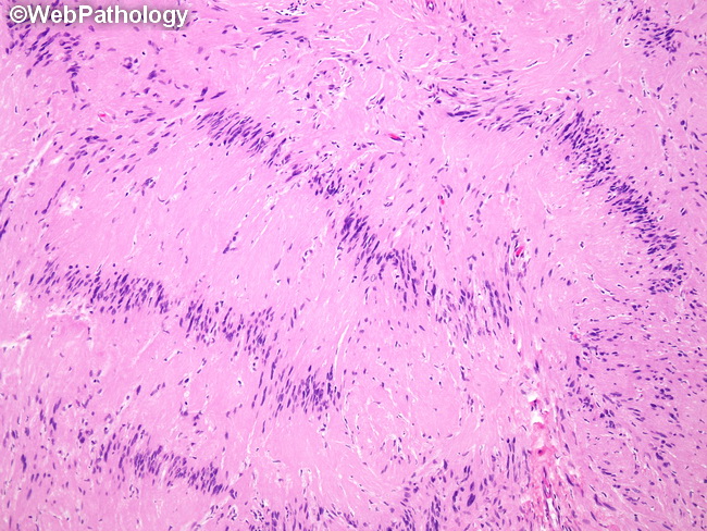
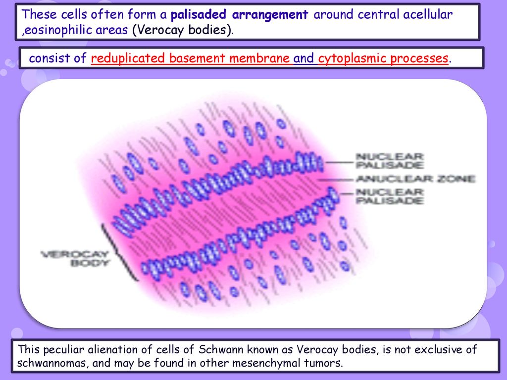
Stanford Pathology on X: "#PathBasics A: Verocay bodies (named after José  Juan Verocay) consist of regions of palisading nuclei that alternate with  an anuclear zone and are classically associated with schwannomas.  #PathTwitter #
Stanford Pathology on X: "#PathBasics A: Verocay bodies (named after José Juan Verocay) consist of regions of palisading nuclei that alternate with an anuclear zone and are classically associated with schwannomas. #PathTwitter #

Verocay body - YouTube

Brain Tumor in Differential Diagnosis of Seizures in Puerperium: Case Report

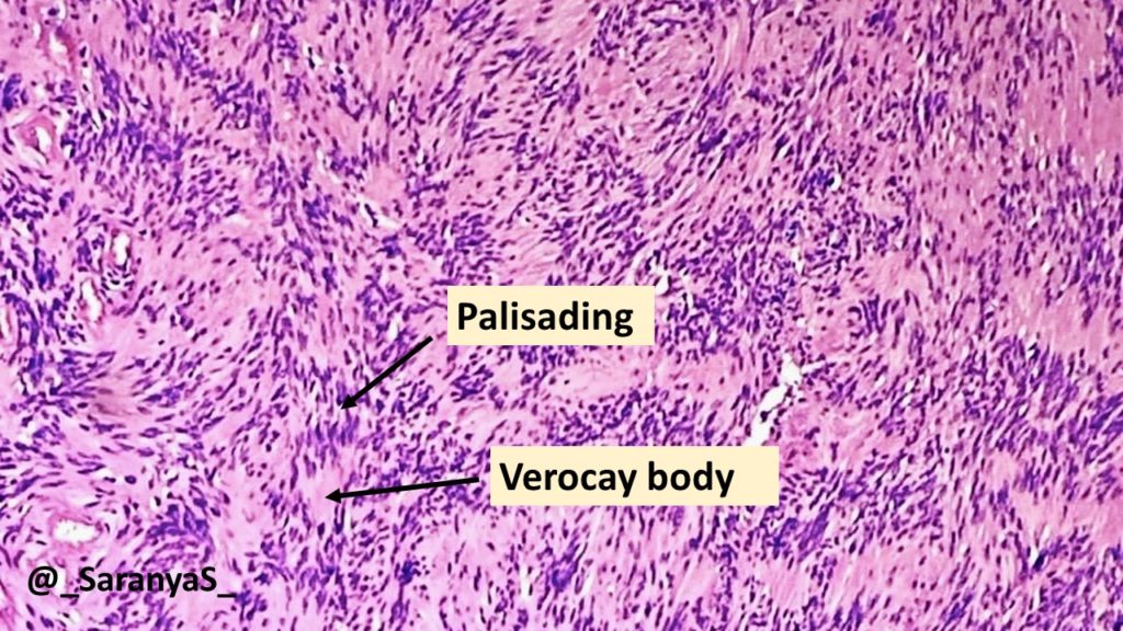
Verocay Bodies in Schwannoma – Adventures in Neuropathology

File:Antoni A area of schwannoma with Verocay bodies.png - Wikimedia Commons

Sanjay Mukhopadhyay on X: "So is a Verocay body Antoni A or B or both?" / X

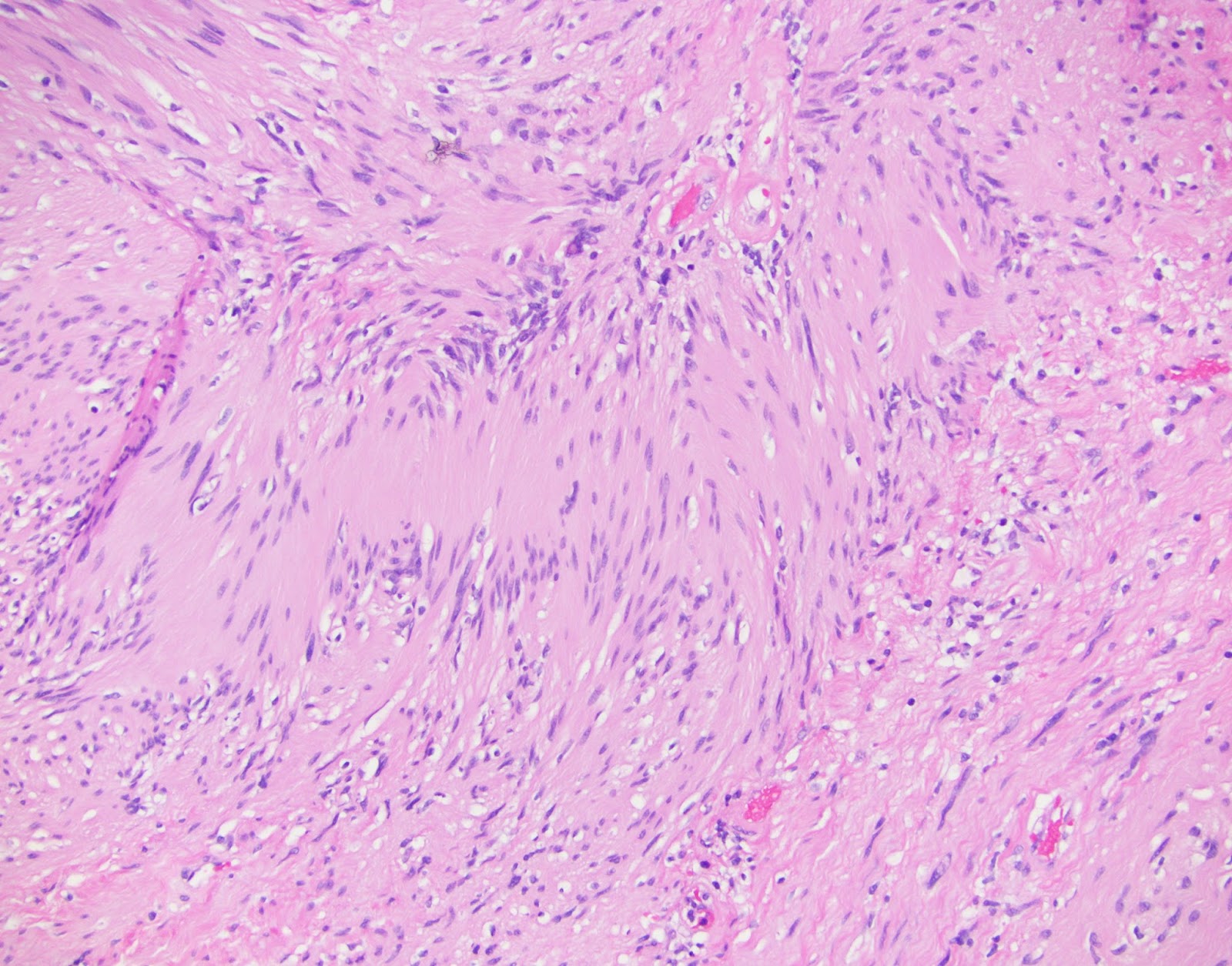
Where are the Verocay bodies?! Recognizing subtle nuclear palisading in  schwannoma. Pathology - YouTube
Where are the Verocay bodies?! Recognizing subtle nuclear palisading in schwannoma. Pathology - YouTube
Final Diagnosis -- Case 868

Verocay body - Wikipedia

Neurilemmoma - Pathology - Orthobullets

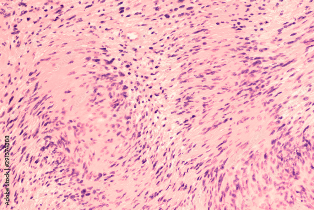
Photomicrograph of a schwannoma, a benign soft tissue tumor of peripheral  nerve sheath, with characteristic nuclear palisading and "Verocay bodies".  Stock Photo | Adobe Stock
Photomicrograph of a schwannoma, a benign soft tissue tumor of peripheral nerve sheath, with characteristic nuclear palisading and "Verocay bodies". Stock Photo | Adobe Stock

Verocay body showing horizontal rows of palisaded nuclei separated by... |  Download Scientific Diagram
Verocay body showing horizontal rows of palisaded nuclei separated by... | Download Scientific Diagram

Webpathology.com: A Collection of Surgical Pathology Images

NEURILEMOMA (SCHWANNOMA) - ppt download

Schwannoma (explained in 5 minutes) Verocay bodies Antoni A & B usmle nerve  sheath tumor pathology - YouTube
Schwannoma (explained in 5 minutes) Verocay bodies Antoni A & B usmle nerve sheath tumor pathology - YouTube
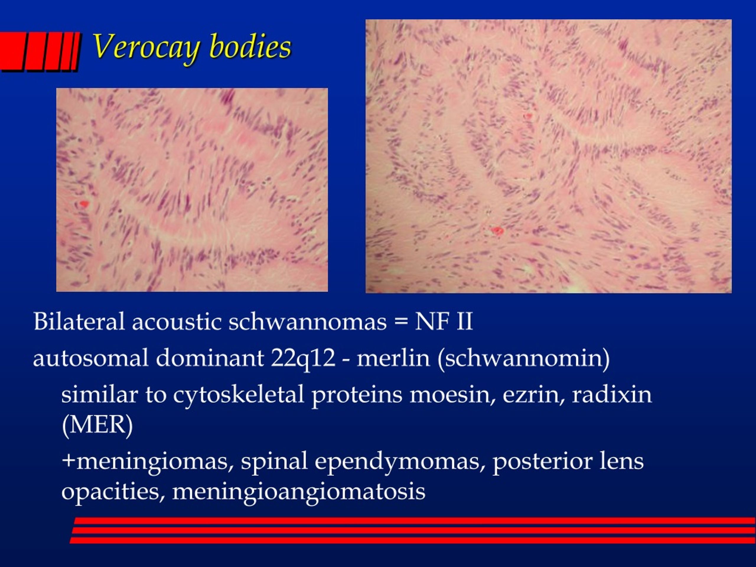
PPT - “SOME BODIES IN THE BRAIN” Noon Diagnostic Conference 11-20-2003  PowerPoint Presentation - ID:208528
PPT - “SOME BODIES IN THE BRAIN” Noon Diagnostic Conference 11-20-2003 PowerPoint Presentation - ID:208528

Pathology of Schwannoma - Pathology Made Simple

Verocay bodies | Radiology Reference Article | Radiopaedia.org

neuropathology blog: Verocay body in a brachial plexus Schwannoma

Schwannoma showing hypodense and hyperdense areas with Verocay bodies (H &  E, 40X) - Indian J Pathol Oncol
Schwannoma showing hypodense and hyperdense areas with Verocay bodies (H & E, 40X) - Indian J Pathol Oncol

Head and Neck Pathology Journal - Well-formed Verocay bodies | Facebook

Schwannoma causing resorption of zygomatic arch – ScienceOpen

PDF] Learning from eponyms: Jose Verocay and Verocay bodies, Antoni A and B  areas, Nils Antoni and Schwannomas | Semantic Scholar
PDF] Learning from eponyms: Jose Verocay and Verocay bodies, Antoni A and B areas, Nils Antoni and Schwannomas | Semantic Scholar